Cover Story
Mutually Interdependent: The Venous and Lymphatic Systems
By Jennifer Boggs | Feature
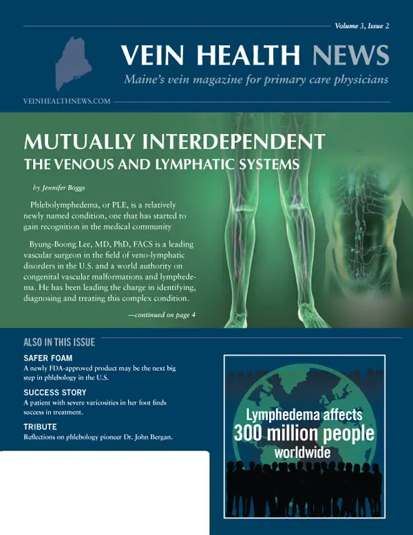
Phlebolymphedema, or PLE, is a relatively newly named condition, one that has started to gain recognition in the medical community. Byung-Boong Lee, MD, PhD, FACS is a leading vascular surgeon in the field of veno-lymphatic disorders in the U.S. and a world authority on congenital vascular malformations and lymphedema. He has been leading the charge in identifying, diagnosing and treating this complex condition.
"For decades, PLE has been unintentionally neglected due to the lack of advanced knowledge of the intertwined venous and lymphatic systems," said Dr. Lee. "All swollen limbs, therefore, should be assessed from a venous as well as a lymphatic point of view."
Edema and lymphedema
Edema is swelling that occurs usually due to the accumulation of excess tissue fluid that had not yet returned to the circulatory system. As healing progresses, the excess fluid leaves the area and the swelling goes down. Edema is a common condition, but its causes are many, including pregnancy, certain medications, congestive heart failure, cirrhosis, kidney disease or damage, chronic venous insufficiency and lymphatic insufficiency are just some of the possible etiologies. It can be generalized, throughout the entire body, or localized in a specific region. It can be temporary or permanent, and come and go, depending upon the underlying condition. Patients will frequently turn to their primary care physician to determine the cause of the swelling, and treatment often means treating the underlying cause of edema.
Lymphedema, on the other hand, occurs when the lymphatic system is damaged or blocked and protein-rich fluid builds up in soft body tissues causing swelling. Clinically, this condition is classified as primary and secondary. Primary lymphedema is caused by the abnormal development of the lymph system, such as congenital malformation; infection or injury in utero; or a side effect to a developmental disorder of the lymphatics. Secondary lymphedema, a more common condition, is caused by damage to the lymph system any time after birth, or by removal of lymph nodes because of injury, trauma, infection of the lymphatics, or cancer biopsy. In fact, lymphedema is a common problem that may be caused by cancer and cancer treatment, although the patient may not notice any swelling until months or years after treatment.
Lymphedema usually affects the arm or leg, but it can also affect other parts of the body. Swelling ranges from mild, hardly noticeable changes in the size of the arm or leg to extreme swelling that can make it impossible to use the affected limb. Unfortunately, there is no cure for this chronic condition, only treatment and management. Treatment modalities for managing the lymphedema include surgery, as well as skin care, compression garments, compression pumps, manual lymphatic drainage, exercises, and most importantly patient education. The combination of treatment interventions is known as Complete Decongestive Therapy (CDT) or Complex Decongestive Therapy in the U.S. and Decongestive Lymphatic Therapy (DLT) internationally.
Two sides of the same coin
For years the medical community has viewed the venous and lymphatic systems as two separate entities: the venous system a series of interconnected blood vessels that return deoxygenated blood from the tissues back to the heart, and the lymphatic system the network of vessels through which lymph drains from the tissues into the blood. A new approach considers the systems two sides of the same coin. To reflect this rapidly developing concept, in 2013 the American Board of Phlebology (the medical specialty focusing on veins) changed its name to the American Board of Vein and Lymphatic Medicine.
According to Dr. Lee, there is no such thing as independent or isolated chronic venous insufficiency (CVI) or chronic lymphatic insufficiency (CLI), especially when the condition remains chronic. "Because these two systems are inseparable and mutually complementary, the failure of one system would burden the other, particularly when one of two systems loses its normal function," said Dr. Lee. "Most of chronic indolent ulcers, for example, are typical of 'secondary' PLE, a combined condition of CVI and CLI."
The presentation of edema in a limb depends upon the relative balance between the insufficiencies of the venous and the lymphatic systems. In the case of CLI, it will be a symptom of the nature of the disorder of the lymphatic system: is it primary, secondary, or a combination of both?. In the presence of CVI, which is characteristic of most venous disorders, the increase in lymph flow becomes much greater than the lymphatic system can transport. In many cases, corrective vein treatment, such as endovenous laser ablation (EVLA), will rectify the venous disorder so that the edema resolves. If the edema persists, however, then the vein specialist or primary care physician should always consider concurrent lymphedema. This diagnosis is integral to proper treatment.
Diagnosing PLE
Since phlebolymphedema is a condition of mixed venous and lymphatic insufficiency, how does one begin to tease it out? According to Dr. Andrea Brennan, OTD, OTR/L, CLT-LANA, DAPWCA, PLE is distinctly different from the low-protein edema observed in pure venous disease. As an occupational therapist with a specialty in lymphedema practice management, Dr. Brennan believes that PLE should be considered in any case of adult onset lower extremity edema. Such consideration ought to lead to a focused physical examination looking for signs of venous insufficiency and appropriate testing so that targeted therapy can be begin.
Changes in the skin are a key way to identify the presence of phlebolymphedema in patients: ankle hyperpigmentation (the darkening of skin) or corona phlebectatica (abnormally dilated veins around the ankle, which can appear as blue or red veins or spots). The distribution of ankle hyperpigmentation in venous disease begins behind, above, or on either side of the ankle, but may, in time, encircle the ankle. Additional hallmarks include toe swelling and pitting edema of the lower limb or dorsum of the foot.
Clinically, the Stemmer sign is a very important component in diagnosing lymphedema. Basically, the Stemmer sign is a test where one pinches or lifts the skin (using the thumb and index finger) over a bony prominence of the foot, such as the base of the second toe. If the skin is difficult to pinch together and cannot be lifted up, or "tented," the Stemmer sign is positive and lymphedema is present. In patients with chronic venous insufficiency, clinical and laboratory evidence will confirm the presence of microangiopathy (a disease of the blood vessels causing them to "leak" protein) of the lymphatic network.
If, based on clinical findings, there is a suspicion of PLE in the swollen limb (with or without ulcers), Dr. Lee recommends an initial evaluation based on a combination of noninvasive tests: "Duplex venous ultrasonography for the venous system and radionuclide lymphoscintigraphy for the lymphatic system should be sufficient to confirm the diagnosis regardless of its etiology." "Further appraisal with invasive tests (ascending and descending phlebography, for example) should be reserved only for a road map when needed for the treatment," he added.
Treatment/management
Compression therapy—bandaging, garments, or both—is the cornerstone of treating any form of lymphedema. Initially, patients are encouraged to wear mild compression for up to 20 hours per day to reduce swelling. Compression stockings and compression alternative devices that apply higher pressure and containment are available and may be more effective in controlling edema.
In very mild cases of phlebolymphedema with a mostly a venous component, Dr. Brennan often recommends multilayer compression wraps or short-stretch compression bandages, so that formal lymphedema treatment may not be necessary. "In these mild cases, the bandages help augment lymphatic flow sufficiently," she said. "As the patient ambulates against bandages, the high sub-bandage pressure can help promote lymph flow through the dermis.". Dr. Brennan will often recommend bandaging the toes with specialized minimal-compression toe bandages to prevent retrograde accumulation of lymph fluid to the toes and distal forefoot.
For patients with any significant degree of phlebolymphedema, the standard of care is Decongestive Lymphatic Therapy by a qualified therapist, including manual lymphatic therapy, skin care, exercise, and short-stretch compression. "The goal of DLT should be to control all the swelling all the time, because anytime there is an accumulation of pro-inflammatory lymph fluid in the interstitium, there is increased risk to the patient of infection and long-term lipodermatosclerotic processes," said Dr. Brennan. Lipodermatosclerosis is an acute or chronic condition that includes skin changes of the lower legs, such has redness, hardening or a "bowling pin" appearance.
PCPs and PLE
All swelling, no matter the cause, is lymphatic overload. Systemic and contributing factors can play a significant role in causing or exacerbating PLE and should be corrected when possible. Dr. Brennan suggests referring patients to a board certified phlebologist to address venous system dysfunction. Additionally, the use of a certified lymphedema therapist, or in close conjunction with wound care treatment, is tantamount to providing quality care. When primary care physicians or other healthcare professionals encounter chronic lymphatic insufficiency, or chronic venous insufficiency in the presence of persistent edema, Dr. Lee advises that they, "always consider these two systems together as a whole before committing to a treatment strategy."
RESOURCES
- Bunke N, Brown K, Bergan J. (2009) Phlebolymphedema: usually unrecognized, often poorly treated. Perspect Vasc Surg Endovasc Ther. 21 (2): 65-68
- Chatham N, Thomas L, Molyneaux, M. Dermatologic difficulties: Skin problems in patients with chronic venous insufficiency and phlebolymphedema. Wound Care Advisor. 2013:2(6):30-36.
- "Don't Forget LYMPHEDEMA!" VEIN Magazine 31 August 2012: 33-38. Print.
- Farrow W. Phlebolymphedema—a common underdiagnosed and undertreated problem in the wound care clinic. J Am Col Certif Wound Spec. 2010;2(1):14-23.
- Lee BB, Andrade M, et al. (2013). Diagnosis and treatment of primary lymphedema. Consensus document of the International Union of Phlebology (IUP)-2013. International Angiology, 32(6), 541-574.
- Piller N. Phlebolymphoedema/chronic venous lymphatic insufficiency: an introduction to strategies for detection, differentiation and treatment. Phlebology. 2009;24(2):51-55.
- Vascular Disease Foundation. Lymphedema. http://vasculardisease.org/flyers/lymphedema-flyer.pdf Accessed June 1, 2014.
Tribute
A Tribute to John J. Bergan, MD
By Dr. Cindy Asbjornsen
Vascular surgeon Dr. John Bergan wrote the book on vein care—well, technically he co-edited it. Considered by most to be the authoritative work on veins and venous treatment, The Vein Book was one of my first textbooks. As a researcher, Dr. Bergan authored 39 books and more than 750 scholarly papers. It's clear through his writing that he was extremely intelligent, diligent and inquisitive, qualities he encouraged in everyone around him.
I met Dr. Bergan in 2005 at my first American College of Phlebology meeting. I walked up to him and introduced myself, and when I did, he immediately asked me a hundred questions about myself, and how I got interested in vein care. I remember he was excited because I was already passionate enough about vein care to fly to San Francisco for a vein meeting, even though I hadn't yet finished my residency. I wanted to learn about veins and he wanted to teach everyone!
Truly a founding father of modern phlebology, Dr. Bergan established the Vein Institute of La Jolla in 2000. He was among the first doctors in the U.S. to use foam sclerotherapy and other noninvasive interventions to treat vein disorders. As an educator, Dr. Bergan taught at UC San Diego School of Medicine, where he was professor of surgery. His research at UCSD (in collaboration with Professor Geert Schmid Shoebein) revealed the fundamental cause of venous reflux. Dr. Bergan also created and directed the nation's first Phlebology Fellowship at the American College of Phlebology, which commenced in July 2007.
Dr. Bergan helped to develop many practice guidelines, including guidelines for venous disease diagnosis and treatment. He served as president of a number of societies, including the American Venous Forum, the Society for Vascular Surgery, and the Lymphedema Association of North America. Dr. Bergan was a great contributor not just to Phlebology, but to a variety of medical fields. He was chief of the Division of Transplantation at Northwestern University Medical School from 1969 to 1976 and chief of the Division of Vascular Surgery from 1976 to 1988.
That's one thing that's always struck me about Dr. Bergan: he was always pushing to further the science behind all medicine. He was a great role model for me as a doctor. And as a human being, the words that come to mind are kind and caring. He made me feel welcome and valued from the minute I met him, to the last time I saw him—at a vein meeting, of course. Dr. Bergan died of complications of a neurological disease on June 11, 2014 at a hospice in Chicago. He was 87. He was a wonderful man, teacher and phlebologist.
Vein Tech
Varithena Moves Vein Care Forward
By Jennifer Boggs
Since the FDA approved radiofrequency endovenous ablation in 1999, the therapies and techniques used to treat venous disease have continued to make great strides. A new product called Varithena, an injectable foam used in sclerotherapy, may be the next big step in modern phlebology.
Sclerotherapy in review
Sclerotherapy is used to treat spider veins and varicose veins by injecting a sclerosing agent into the interior of the troublesome vein. This substance causes the vein walls to become sticky and seal shut. The collapsed vein is reabsorbed into local tissue and eventually disappears. There is little risk of complication, and patients often experience an immediate relief of symptoms.
Light-assisted sclerotherapy is used to treat smaller veins below the skin's surface. These reticular veins are responsible for feeding the veins that are visible on the surface of the skin. A small, powerful light illuminates the veins and tissue directly below the patient's skin, allowing the physician to clearly identify the source of the venous dysfunction and to perform the procedure. Ultrasound-guided sclerotherapy is used for larger, less superficial veins that cannot be seen with transcutaneous illumination (light used to view veins near the surface). Due to the proximity of some of these veins to nerves, arteries and other structures, a skilled ultrasound sonographer is critical to the success of this procedure. After sclerotherapy, treated veins often fade within a few weeks, although it may take longer to see the full results. In some instances, several sclerotherapy treatments may be necessary.
Liquid vs. foam
There are several kinds of liquid sclerosing agents. The most popular modern sclerosant is called polidocanol. Polidocanol is a detergent by nature and, therefore, can form bubbles easily. Foam sclerotherapy is, simply put, the mixture of the liquid sclerosant with a gas to create those bubbles. Phlebologist Dr. Kenneth L. Todd III, lead physician and founder of the Southeast Vein and Laser Center in Dothan, Alabama, described foam as "a solution comprised of millions of tiny bubbles compressed together." When the foam is injected into the veins, the micro-bubbles touch the vein walls and destroy them (the object of sclerotherapy).
In the treatment of varicose vein treatments, foaming the sclerosant enables it to displace the blood in larger veins, rather than just mixing with it, as a liquid sclerosant would. That means foam provides longer contact time between the chemical and the vein wall and is more effective on larger veins, such as the great saphenous vein (GSV) and other truncal veins. Liquid sclerosants work best on smaller, reticular veins. Another significant advantage of foam, according to Dr. Todd, is that it is echogenic, which means that it's easily visible on ultrasound, increasing the accuracy with which individual veins can be treated. In addition, because the efficacy of the sclerosing agent is increased with foam, a lower concentration can be given to treat varicose veins, which means using a smaller total dose of sclerosant to achieve the desired effect.
The most common method for producing foam for sclerotherapy is the "Tessari technique," which involves manually mixing a small volume of liquid sclerosant with a volume of room air or other gas using two syringes and a three-way tap, or stopcock. While physician-compounded, non-proprietary foam is inexpensive to make, experts caution that there are risks: there have been a number of published cases of air emboli (small bubbles of gas in the bloodstream) from so-called "homemade foam" leading to neurological events in patients, including strokes. No physician-compounded foam is approved by the FDA.
Standardized bubbles
Vein care specialists in the Europe and U.K. have been using physician-compounded foams to treat varicose veins for at least ten years. In the U.S., however, a proprietary foam called Varithena was approved by the FDA in the fall of 2013. According to Dr. Robert Diamond, Vice President of Medical Affairs at BTG, the international specialist healthcare company that manufactures Varithena, it is a unique proprietary mixture of one percent polidocanol, 65% oxygen, 35% carbon dioxide, and less than .8% of nitrogen.
"When a physician creates his own foam, he uses room air, which can contain as much as 78% nitrogen—compared to Varithena, which has extremely low nitrogen," explained Dr. Diamond. "While oxygen and carbon dioxide are rapidly absorbed into the blood stream, nitrogen is very slowly absorbed and those bubbles can persist and possibly cause a gas embolism and obstruct an important blood vessel." Perhaps the most important safety feature of Varithena is the consistency of its bubbles: each one is between 100-500 microns. With its consistently uniform density, stability, and range of bubble size, Varithena it is much more predictable for vein doctors to use on their patients than physician-compounded foam, which isn't subject to the same manufacturing standards.
Results and risks
Varithena has been studied in more than 1,300 patients in a series of phased clinical trials, and the product has FDA approval for the treatment of the GSV, saphenous accessories, and varicose tributaries above and below the knee. Data from BTG reported that their studies "achieved primary, secondary and tertiary efficacy endpoints in patients with vein diameters ranging from 1.5 to 25.9 mm." In other words, significant improvement was noted in symptoms and appearance by both the patients and their physicians, and the treated vein closed more than 80% of the time with only one treatment.
The most common adverse event recorded was pain or discomfort at the injection site. As with other varicose vein treatments, Varithena can cause deep venous thrombosis under specific conditions. Dr. Diamond explained that, "with any varicose vein treatment, there is always the risk of DVT, because the veins that are being treated are connected to the deep venous system.". Another risk, though extremely rare, is allergy to polidocanol itself.
Varithena will be available for administration by trained physicians in the third quarter of 2014. As with any new therapy, price, as well as insurance codes and coverage, will be evolving as the product becomes commercially available. Physicians interested in learning about Varithena should contact BTG. Varithena may not totally replace other venous treatments, such as laser ablations, or even sclerotherapy using liquid sclerosants, but the mode of therapy really depends on the patient and his or her goals. "Every time a phlebologist sees a patient, he has to assess what's best for that patient's anatomy," said Dr. Todd. "We make that call for every individual patient."
FAVQ
Pronounced Foot Veins
By Dr. Cindy Asbjornsen
The last issue of Vein Health News (Volume 3, Issue 1) featured a cover story about veins in relation to feet. In the story we included a photo of a woman's foot with a large, ropy vein winding its way up her ankle. That foot belongs to one of my patients, and several people have asked me if she received treatment for that pronounced vein. The answer is yes! This 45 year-old patient came to see me complaining of aching, cramping, pain, heaviness and numbness in her legs bilaterally. All of her symptoms worsened with prolonged standing, and she felt self-conscious about wearing clothes that showed her legs. She would often have difficulty falling asleep because her legs bothered her greatly.
The onset of her varicosities was approximately 23 years earlier when she was pregnant with her son. After an initial exam, including ultrasound mapping, I diagnosed her with insufficiency of the great saphenous veins bilaterally. I treated her with bilateral endovenous laser ablations (EVLA) to her great saphenous veins, as well as multiple sessions of sclerotherapy to clean up the surrounding veins. The outcome for this patient was a full resolution for all symptoms, including the bulging veins. The patient enjoyed wearing shorts and skirts this summer and is happy to be free of her previous symptoms.
Patient Perspective
One Patient's Perspective: Proactive Prevention
By Benjamin Lee
Joe Knapp of Jay, Maine was 14 years old when he was hit head-on by a car while he was riding a bicycle. The accident fractured his left femur at the proximal one-third of the bone and put him in traction for a week. Femur rodding was a new procedure at the time, but the bone healed quickly and Knapp was able to resume sports like basketball and cross-country running. Fast-forward about ten years. Knapp started to see varicose veins gradually forming on his left ankle and along the scar from the rodding surgery he'd had in high school. At 35, he started to wear knee-high support hose to protect his legs from long shifts as a nurse on a medical-surgical unit. But his leg got worse.
"I wore support hose for more than a decade, but as the years went on they were less and less helpful," said Knapp. "When I turned 50, they stopped being effective at all, and I began to notice an itching and heaviness in my legs." Fortunately, Knapp had some training in wound care and knew that all venous ulcers start somewhere, often with symptoms of itching and swelling. In addition, there was a history of lymphedema in Knapp's family. His mother had Milroy's disease, a condition characterized by lymphedema in the legs and caused by congenital abnormalities in the lymphatic system. Knapp's sister also developed swollen ankles in her thirties, and he also has ankles that swell easily.
Knapp's family history of lymphedema, combined with years of working on hospital cement tile floors, motivated him to investigate further. And his 2½ years of wound care convinced him to do something about it before it got worse. "I've seen what can happen when some people don't do anything to address the problem," said Knapp. "The varicose vein can eventually become a venous ulcer, a wound—and wound healing can turn into a long, arduous process." He added that once the tissue "has had an assault on the system," it becomes more difficult to repair as the person ages. The presence of varicosities, or even swelling, can exacerbate it.
In Knapp's case, awareness led to action. He went to a board-certified phlebologist for a physical exam and diagnostic ultrasound evaluation. Together they decided to pursue sclerotherapy, a procedure that Knapp described as "smooth and virtually painless.". He had five sclerotherapy treatments on his legs and the results were immediately apparent. His legs stopped itching and felt lighter, and according to Knapp "they looked much better too."
Now, three years later, Knapp is a nurse in private practice. Because of his vein experience and the care he received, he expresses his concern to the doctor whenever he sees varicose veins or other venous issues, in an effort to treat the problem early and prevent further problems, such as venous ulcers, infections, superficial thromboses and potentially bleeding. "Just the idea that I could get an ulcer before I turn 60, no thanks!" Knapp stressed. "These kinds of problems don't go away on their own—you need to do something about it."
Concerned about your vein health?
Contact the Vein Healthcare Center for an evaluation.
Contact Us